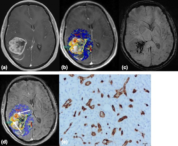
Figure 3.

Images(a-d) of a 44-year-old woman with right temporal anaplastic oligodendrogliomas. (a) Contrast-enhanced T1-weighted image shows a mass with irregular enhancement. (b) Ktrans map shows high Ktrans values in the tumor, including (c) a maximum degree of ITSS in the SWI. (d) Co-registered image of Ktrans and SWI shows that regions of the highest value of Ktrans(arrows) does not correspond with areas of attenuated prominent ITSS in the same segment. (e) Representative immunohistochemical staining(CD34, Original magnification,×200) shows abundant angiogenesis in the tumor, with high MVD, bizarre vascular formation and large VD.
