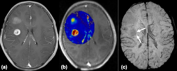
Figure 4.

Images of a 13-year-old boy with left frontal glioblastoma. (a) The contrast-enhanced axial T1-weighted image shows a mass with regular peripheral rim enhancement. (b) The high Ktrans values within the tumor indicates a high permeability of microvessels. (c) However, SWI reveals no evidence of ITSS (arrows).
