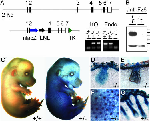
Fig. 1.
Targeted mutation of the Fz6 gene and expression of Fz6-nlacZ in skin and hair follicles. (A Upper) Structure of the Fz6 locus. Black rectangles represent the seven exons; in exons 2–7, the shaded areas represent the Fz6 coding region. (Lower Left) Targeting vector. LNL, PGK-neo gene flanked by loxP sites; TK, herpes simplex virus thymidine kinase cassette. (Right) Genotyping of tail DNA with PCR primers specific for the neo-Fz6 junction (KO) or endogenous Fz6 sequences deleted in the targeted allele (Endo). (B) Immunoblot of proteins from P1 skin probed with affinity-purified antibodies directed against the C-terminal 17 aa of Fz6. A band of ≈85 kDa is present in the Fz6(+/+) sample and absent in the Fz6(-/-) sample and is presumed to be Fz6; a crossreacting band of ≈55 kDa is seen in both samples. Protein molecular mass standards from top to bottom are 100, 90, 80, 70, 60, and 50 kDa. (C) X-Gal stain of E14.5 embryos. (Left) Fz6(+/+). (Right) Fz6(+/-). (D and E) X-Gal stain of Fz6(-/-) skin at E16.5; in E, a similar section has also been immunostained for keratin-14 (brown). X-Gal staining is present in the periderm, epidermis, and hair follicles. (F) Enlarged image of the flank of the Fz6(+/-) embryo in C shows X-Gal stain concentrated in developing hair follicles. (G) X-Gal stain of Fz6(+/-) skin at ≈P1. C–G show strong expression of the Fz6-nlacZ reporter in developing epidermis and hair follicles. Fz6(+/-) and Fz6(-/-) show the same pattern of X-Gal staining.

