Fig. 3.
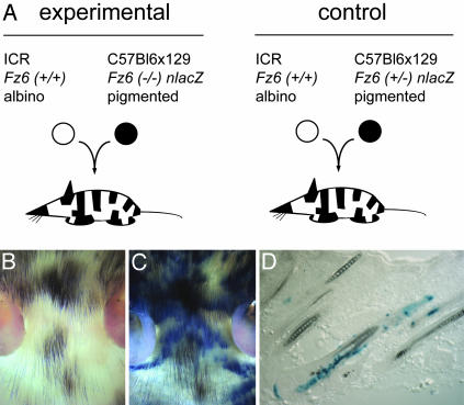
Generation of chimeric mice. (A) Diagram of the experimental (Left) and control (Right) embryo aggregation protocols. (B and C) Unstained (B) and X-Gal-stained (C) flatmounts of skin from the head of an albino ICR Fz6(+/+):pigmented C57BL6 × 129 Fz6(-/-) chimeric mouse at approximately P10 shows the independent patterns of tissue chimerism for epidermal cells (X-Gal-stained or unstained) and melanocytes (pigmented or albino). A small amount of tissue shrinkage accompanies the fixation and X-Gal staining procedure. (D) An X-Gal-stained frozen section from the abdomen of a chimeric mouse shows the characteristic heterogeneity of Fz6(-/-) epidermal cells (X-Gal-stained) within single hair follicles. This image also shows, on the scale of individual hair follicles, the independent patterns of tissue chimerism for epidermal cells and melanocytes.

