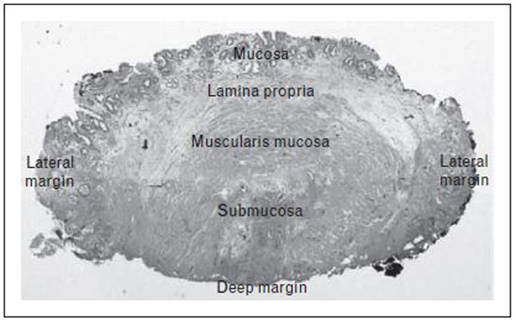
FIGURE 1. Endoscopic Mucosal Resection Histopathology.

Histopathology of cross-section of endoscopic mucosal resection specimen showing mucosa, lamina propria, muscularis mucosa and submucosa. Focal high-grade dysplasia within the mucosal layer with clear deep and lateral margins.
