
Fig. 2.
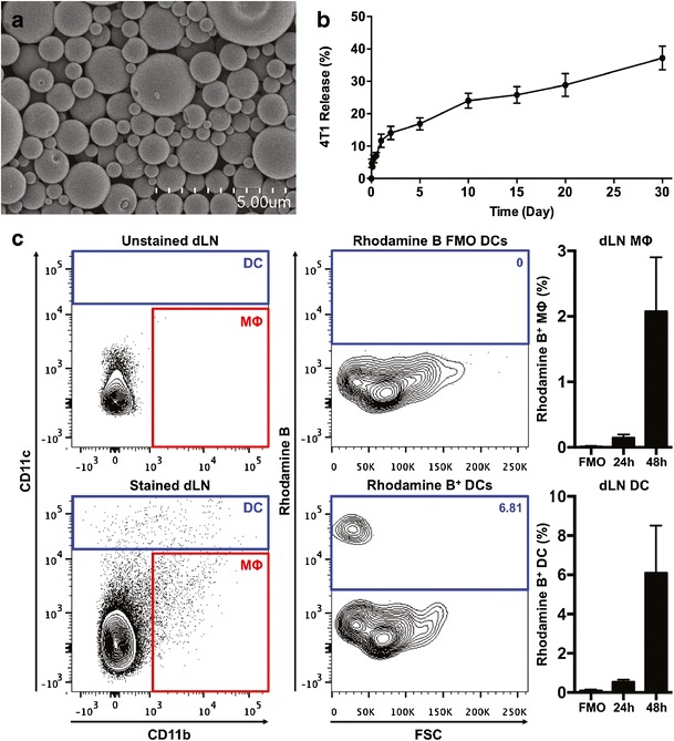
PLGA MPs are efficiently phagocytosed by DCs and MΦs in vivo. a Scanning electron photomicrograph of 4T1-loaded PLGA MP. b 4T1 lysate release profile from PLGA MPs cultured in PBS at 37°C. Data from three batches of 4T1 MPs. Mean ± S.E.M. c In vivo trafficking of Rhodamine B-loaded PLGA MP. Left: Representative flow cytometry plots indicating the gating strategy for DC and MΦ populations in the vaccine-dLN (inguinal). Center: Rhodamine B gating defined using FMO controls. Right: Frequency of Rhodamine B-positive phagocytes in the dLN 24 and 48 h after MP injection. Data shown (mean ± S.E.M.) are pooled from two experiments using a total of eight mice/time point