
Figure 7.
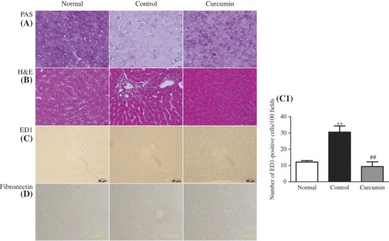
Effect of curcumin on histopathological changes. Histological staining with PAS in liver (A) shows that glycogen contents of rat liver decreased in diabetic animals when compared with those of normal control animals, but these levels increased to near normal after treatment with curcumin. In H&E, (B) light microscopic photographs of livers of experimental animal showed the liver of normal control group, lipid accumulation indicated by the unstained area in liver tissues, microvascular fattening and focal necrosis, and portal inflammation in the untreated diabetic group; in curcumin-treated diabetic group, the severity of these changes was less than those in the untreated diabetic group. (C and C1) Immunohistochemical staining for macrophage (ED1-positive cells) and its quantification graph in each group. (D) Immunohistochemical staining for fibronectin in liver section. Each bar represents mean ± SE. Normal, age-matched normal rats; Control, untreated diabetic rats; Curcumin, diabetic rats treated with curcumin 100 mg/kg/day. ∗∗p < 0.01 versus Normal, ##p < 0.01 versus Control.