Figure 1.
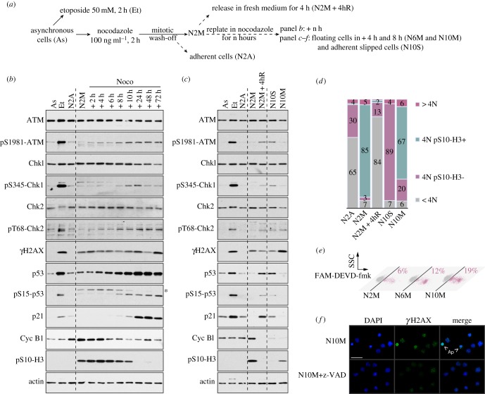
The period of mitotic arrest controls subsequent DNA damage signalling, cell cycle progression and apoptosis. (a) Experimental protocol. (b) DNA damage signalling in response to a prolonged mitotic arrest. Samples were analysed by immunoblotting using the antibodies indicated. Asterisk denotes a non-specific signal on pS15-p53 blot. (c,d) DNA damage signalling in G1 following mitotic arrest. Samples were analysed by immunoblotting using specified antibodies (c), and cell cycle profiles were characterized by flow cytometry using propidium iodide as a marker of DNA content and phosphorylated Ser10 histone H3 as a marker of mitosis (d). (e) Induction of apoptosis during a mitotic arrest. Cells were incubated with a FAM-DEVD-fmk probe and analysed by flow cytometry. Percentages represent the amount of active cleaved caspase-3/7 positive cells. (f) γH2AX induction in cells arrested in mitosis is caspase-dependent. Cells were synchronized for 10 h in mitosis in the presence of z-VAD-fmk where indicated, cytospun and immunostained using an anti-γH2AX antibody. Representative microscopic fields are shown; Ap, apoptotic cells; scale bar, 50 µm.

