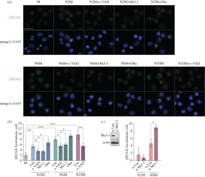
Figure 3.
Mitotic arrest elicits a localized caspase-dependent DNA damage response under the control of Bcl-2 family proteins. Cells were synchronized in prolonged mitosis for different times (N2M, N6M, N10M) and compared with untreated mitotic cells (M). Some cells were co-treated with the pan-caspase inhibitor z-VAD-fmk (20 µM) or the Mcl-1/Bcl-2/Bcl-xL inhibitor Obatoclax (500 nM). U2OS cells over-expressing Mcl-1 were prepared by transient transfection. Cells were cytospun and immunostained using an anti-γH2AX antibody. (a) Representative microscopic fields are shown; scale bar, 40 µm. (b,c) The histograms show the numbers of γH2AX foci per mitotic cell treated as indicated in (a) or in which Mcl-1 was depleted by siRNA (si Mcl-1) compared with control cells in which an irrelevant luciferase siRNA was transfected (si Luc); values are means ± s.d. (n ≥ 3). Statistical differences were analysed using one-way ANOVA statistical tests; *p < 0.05, **p < 0.01 and ***p < 0.001.

