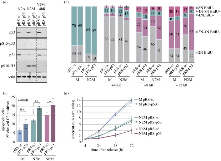
Figure 5.
The role of p53 in the fate of cells following a mitotic arrest. (a) Characterization of p53-depleted U2OS cells. Stably transfected U2OS cells were treated as in figure 1a and analysed by immunoblotting using the specified antibodies. (b) Role of p53 in cell cycle progression following a mitotic arrest. Stably transfected U2OS cells treated as indicated were analysed by flow cytometry. The cumulative histograms show the percentage of cells in the different phases of the cell cycle, according to BrdU incorporation and DNA content. Data shown are from a representative experiment repeated three times. (c,d) Role of p53 in the viability and proliferation of cells following a mitotic arrest. (c) Cells treated as depicted in (a) were incubated with an FAM-DEVD-fmk probe and analysed by flow cytometry. The percentage of apoptotic cells with active caspase 3/7 is shown, values are means ± s.d. (n = 3). Statistical differences were analysed with the Mann–Whitney test; n.s., non-significant, *p < 0.05, **p < 0.01. (d) The relative number of viable, adherent cells was determined by following cell proliferation using IncuCyte-based cell density analyses. Values are means ± s.d. from quadruplicate wells of a representative experiment repeated three times.

