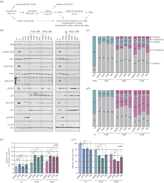
Figure 6.
Inhibition of DNA damage response kinases after release from mitotic arrest enhances the effects of microtubule poisons. (a) Experimental protocol. (b) Selective inhibition of PIKKs affects DNA damage signalling after a mitotic arrest. Samples were analysed by immunoblotting using the specified antibodies; asterisk denotes a non-specific signal on pS15-p53 blot. (c,d) Inhibition of PIKKs affects cell cycle progression following mitotic arrest. Cells arrested in mitosis for 2 h with nocodazole (c) or normal mitotic cells (d) were released as depicted in (a) and analysed using flow cytometry. The cumulative histograms show the percentage of cells in the different phases of the cell cycle, according to BrdU incorporation and DNA content. Data shown are from a representative experiment (n ≥ 3). (e,f) Inhibition of PIKKs reduces the viability and proliferation of cells following a mitotic arrest. (e) Cells treated as indicated in (a) were incubated with an FAM-DEVD-fmk probe to identify apoptotic cells and analysed by flow cytometry. The percentage of apoptotic cells is shown, values are means ± s.d. (n ≥ 3). Statistical differences were analysed with the Mann–Whitney test; n.s., non-significant, *p < 0.05, **p < 0.01 and ***p < 0.001. (f) The relative number of viable, adherent cells was determined by crystal violet assay. Values are means ± s.d. from quadruplicate wells of a representative experiment (n ≥ 3).

