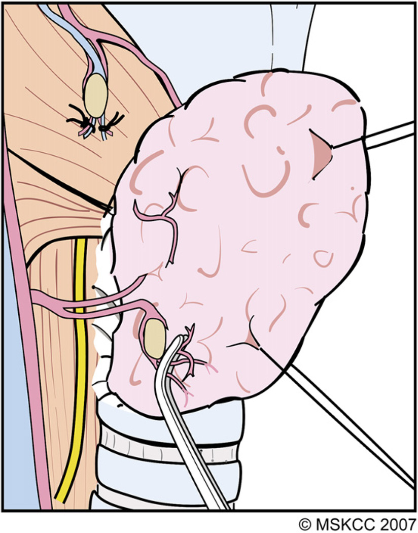
Fig. 8.
The terminal branches of the ITA that are encountered distal to the PT gland after they have supplied these glands are divided and ligated carefully. As the areolar tissue around the thyroid capsule is peeled away along with the PT gland, the RLN comes into view. (Courtesy of the Memorial Sloan-Kettering Cancer Center, New York, NY; with permission.)

