Figure 6.

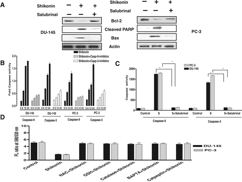
ER stress inhibition attenuates Shikonin induced mitochondrial apoptosis. (A) DU-145 and PC-3 cells were treated with either Shikonin (2.5 μM) alone for 24 h or pretreated with ER stress inhibitor Salubrinal (10 μM) for 24 h and subsequently treated with Shikonin. Protein expression was measured by western blotting as described in the Methods section. Shikonin treatment resulted in a marked reduction in the expression of Bcl-2 and an increase in the levels of Bax and cleaved PARP when compared with the controls. Pretreatment with ER stress inhibitor Salubrinal led to the increased expression of Bcl-2 and the decreased expression of Bax and cleaved PARP. (B) Pretreatment with caspase inhibitors reversed Shikonin induced caspase activation. Treatment of cells with Shikonin (2.5 μM) for 24 h resulted in a marked reduction of shikonin induced caspase-9 and caspase-3 activities. (C) Inhibition of ER suppressed shikonin induced casapase activity. Prostate cancer cells were pretreated with Salubrinal (10 μM), caspase 3 and 9 activity were performed by colorimetric assay as described in material and method section. (D) ER stress regulates mitochondrial membrane potential in Shikonin treated DU-145 and PC-3 cells. DU-145 and PC-3 cells were treated with either Shikonin (2.5 μΜ) alone or along with pretreatment with salubrinal (10 μM) for 24 h, and then incubated with mitochondria specific dye JC-1 (10 m g/ml aft 37°C for 15 min), and the florescence was monitored using fluorimeter (Excitation 530 nm/Fluorescence 590 nm) for measurement of the mitochondrial membrane potential. Shikonin treatment significantly reduced mitochondrial membrane potential whereas pretreatment of ER stress inhibitor reversed this effect. Data is expressed in means ± SEM and represents the results of three independent experiments.
