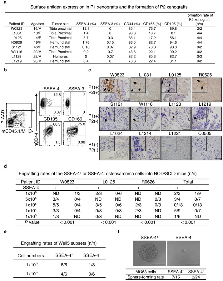
Figure 1. SSEA-4 Labels Xenografting-TICs Present in a Minority of Human Osteosarcoma Cases.
(a) The frequency of SSEA-4+ cells in P1 xenografts correlates with xenograft-formation potential in secondary recipients. (b) Live 7-AAD− and murine CD45+/MHC-I+-excluded osteosarcoma xenograft cells were analyzed for expression of the indicated antigens by flow cytometry. (c) Primary osteosarcoma samples that generate both P1 and P2 xenografts show higher SSEA-4-staining intensity than those that produced only P1 xenografts. Primary specimens that produced no P1 xenografts stained negatively for SSEA-4. Immunohistochemical staining signals for SSEA-4 are indicated by arrows. Scale bar represents 100 μm. (d) Engrafting efficiencies of SSEA-4+ or SSEA-4− cells freshly isolated from three individual tumorigenic xenografts (P2 to P5). (e) Xenografting efficiency of SSEA-4+ or SSEA-4− osteosarcoma cells isolated from in vitro-cultivated Well5 cells (P < 0.05). (f) Sphere-forming rate of SSEA-4+ or SSEA-4− cells isolated from in vitro cultivated osteosarcoma MG63 cells (P < 0.05). Microscopic inspection of representative tumor-sphere or cellular debris is shown in the upper panel.

