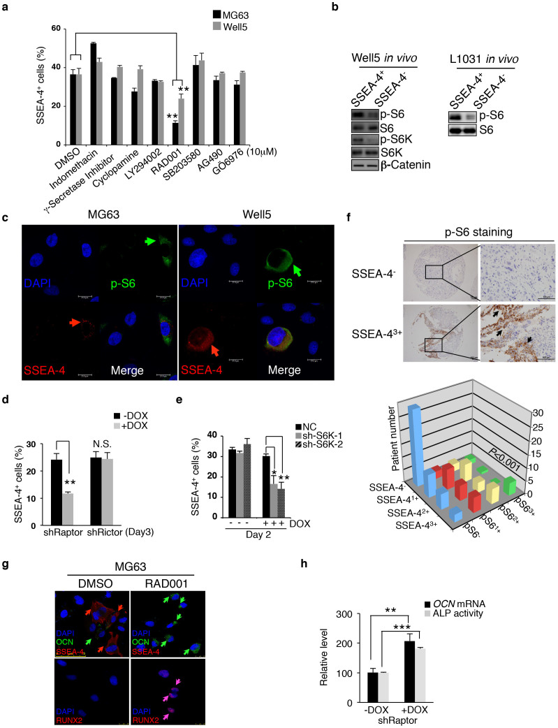
Figure 4. mTORC1 Activity Maintains SSEA-4+ TIC Frequency.
(a) MG63 or Well5 cells were treated with negative control DMSO, Wnt-β catenin inhibitor indomethacin, Notch inhibitor γ-secretase inhibitor, Hedgehog inhibitor cyclopamine, PI3K-AKT inhibitor LY294002, mTOR inhibitor RAD001, p38 MAPK inhibitor SB203580, JAK-STAT inhibitor AG490, or PKC inhibitor GO6976 for 24 hours and the frequency of SSEA-4+ cells was measured by flow cytometry. Results are expressed as the mean ± SD (**P < 0.01). (b) Western blotting assays for phosphorylated levels of mTORC1 pathway components S6K or/and S6 as well as the β-catenin level in SSEA-4+ or SSEA-4− cells freshly sorted from osteosarcoma xenografts. The cropped blots were run under the same experimental conditions. The full-length blots can be found in Supplementary Figure 8. (c) Immunofluorescent co-staining of SSEA-4 and p-S6 in cytospun MG63 and Well5 cells. (d–e) The effects of Dox-inducible Raptor or Rictor knockdown (d) or S6K knockdown (e) on the frequency of SSEA-4+ cells in MG63 cell culture. Results are expressed as mean ± SD (*P < 0.05, **P < 0.01). NC: control shRNA; shRaptor: shRNA for Raptor; shRictor: shRNA for Rictor; sh-S6K-1 and sh-S6K-2: shRNAs for S6K. (f) p-S6 level is positively correlated with SSEA-4 staining intensity among 98 human osteosarcoma samples (P = 0.000155). Representative immunohistochemical staining of p-S6 in one SSEA-4− and SSEA-43+ sample is shown on the left panel. Scale bars represent 100 μm. (g) MG63 cells were treated with DMSO or RAD001 for 3 days, then stained with the fluorescent antibodies against SSEA-4, OCN and RUNX2, and viewed microscopically. (h) ALP activity and OCN mRNA levels were measured in MG63 cells with or without Raptor knockdown (**P < 0.01, ***P < 0.001).

