Figure 4.

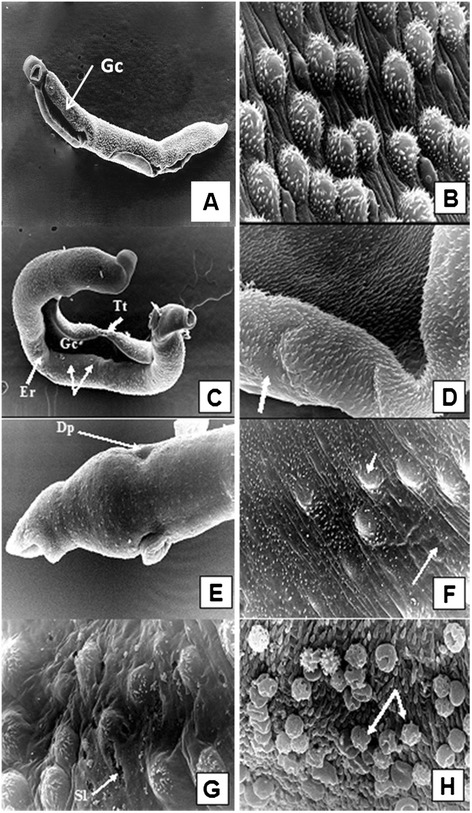
Scanning electron micrographs of adult S. mansoni male worms recovered from unimmunized group (A and B) and from FSm14/29-immunized group (C-H). (A) A male worm obtained from infected control group showing normal body configuration, typical oral and ventral suckers, and distinct tegumental structure with evident normal spines on the dorsal surface behind the beginning of the gynecophoric canal (Gc), X200. (B) Tegument of the middle dorsolateral region showing numerous large tubercles, each bearing numerous spines. Intertubercular ridges and ciliated sensory papillae are obvious, X 5,000. (C) Tegumental surface alterations in adult male from infected FSm14/29-immunized group demonstrating widening of the gynecophoric canal (Gc), loss of some areas of the tegumental tubercles and ridges (arrows), surface erosions (Er), tegumental tear (Tt), and bossing of the terminal end of the worm, X200. The posterior end shows several areas of bulging. (D) Higher magnification of a conspicuously widened gynecophoric canal, along with restricted spine loss (arrow), X5,000. (E) Swollen area between the oral and ventral suckers showing circumferentially arranged ridges with sensory papillae rows. An evident dorsal dimple is witnessed (Dp), along with suckers oedema and blebbing, X2,000. (F) Eminent flattening of the dorsal tubercles along with shortened spines which were irregularly positioned or even lost in some areas (arrows), X5,000. (G) Altered dorsal tubercles revealed peeling and focal sloughing (Sl) of their tegument. Tubercular and inter-tubercular coarse bumpy tegumental irregularities were observed, with occasional burst of some blistered tubercles, X5,000. (H) Host leucocytic (arrows) attachment to the worm surface with evident spine blunting, X7500.
