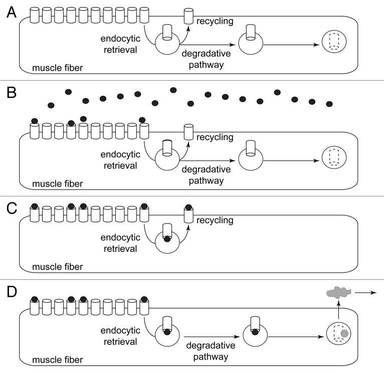
Figure 1. Radioiodine labeling detects total amount of 125I contained in muscle. Scheme illustrating the working principle of the radioiodine approach. (A) Pre-pulse situation. CHRN channels (white barrels) are present at high amounts in the postsynaptic membranes of muscle fibers. Small amounts of CHRN are constantly internalized by endocytic retrieval. From there, CHRN might undergo recycling back to the plasma membrane or follow a degradative pathway. (B) Pulse labeling with 125I-BGT (black dots). About 10% of the 125I-BGT binds to CHRN in the injected hind limb, the rest of the 25I-BGT is washed out and binds to other muscles or is excreted. (C and D) CHRN labeled with 125I-BGT undergo either recycling (C) or are degraded (D). Only in the latter case, 125I is removed from the muscle (gray cloud), i.e., the radioiodine approach measures the rate of 125I removal from the muscle as a consequence of CHRN degradation.

An official website of the United States government
Here's how you know
Official websites use .gov
A
.gov website belongs to an official
government organization in the United States.
Secure .gov websites use HTTPS
A lock (
) or https:// means you've safely
connected to the .gov website. Share sensitive
information only on official, secure websites.
