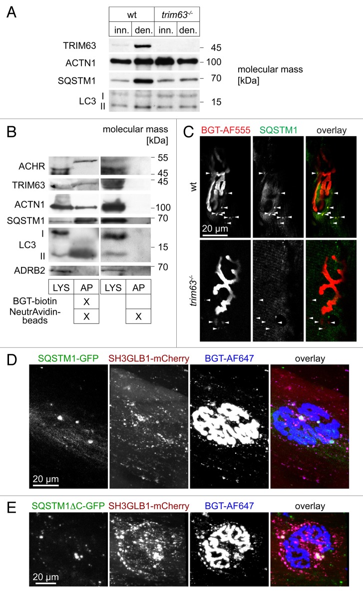
Figure 8. SQSTM1 is upregulated in a TRIM63-dependent manner, it colocalizes with endocytic CHRN and regulates CHRN carrier formation. (A) Representative western blot signals against TRIM63, ACTN1 (loading control), SQSTM1 and MAP1LC3A from lysates of WT or trim63−/− gastrocnemius muscles, that were either innervated (inn.) or denervated for 5 d (den.). (B) WT mouse gastrocnemius muscles were injected with BGT-biotin (left 2 lanes) or with saline (right 2 lanes). Five hours later, mice were sacrificed, and muscles harvested and lysed. Subsequently, BGT-biotin labeled CHRN were sedimented with NeutrAvidin-coupled beads. Depicted are representative western blot immunosignals using antibodies against CHRN, SQSTM1, MAP1LC3A, TRIM63, ACTN1 (positive control), and ADRB2 (negative control). LYS, lysate; AP, biotin-neutravidin affinity precipitate. (C) Denervated EDL muscles from WT and trim63−/− mice were sectioned and stained with BGT-AF555 against CHRN and with an antibody against SQSTM1. Shown are single confocal sections depicting parts of individual NMJs surrounded by some punctate CHRN-positive structures (arrowheads). While in the WT most of these puncta also exhibit SQSTM1 immunofluorescence signals, this is not the case in muscle lacking TRIM63. In the overlay pictures, BGT and anti-SQSTM1 signals appear in red and green, respectively. (D and E) WT tibialis anterior muscles were cotransfected with SH3GLB-mCherry and either SQSTM1-GFP (C) or SQSTM1ΔC-GFP (D). After 7 d, BGT-AF647 was injected and 24 h later, muscles were imaged in vivo. Shown are en face maximum z-projections. In the overlay pictures, GFP-, mCherry- and BGT-signals appear in green, red, and blue, respectively.

An official website of the United States government
Here's how you know
Official websites use .gov
A
.gov website belongs to an official
government organization in the United States.
Secure .gov websites use HTTPS
A lock (
) or https:// means you've safely
connected to the .gov website. Share sensitive
information only on official, secure websites.
