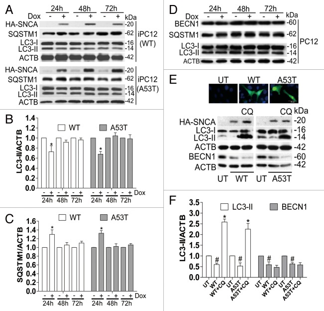
Figure 2. WT and SNCAA53T overexpression inhibits starvation-activated autophagy in a time-course-dependent manner. (A) iPC12 cells were treated with 2 μg/ml Dox for 24, 48, and 72 h respectively and then starved by Earle's balanced salt solution (EBSS) treatment for 2 h. The expressions of LC3-II and SQSTM1 (p62) were determined by western blotting. (B and C) Relative intensity is normalized to that of ACTB. Data are presented as the mean ± SD from 3 independent experiments. *P < 0.05 vs. uninduced control at the corresponding time points. (D) Normal PC12 cells were treated with 2 μg/ml Dox for 24 h. The expressions of LC3-II, SQSTM1 and BECN1 were determined by western blotting. Experiments were performed 3 times with similar results and the representative blots were shown. (E) The expressions of LC3-II and BECN1 in PC12 cells constitutively expressing GFP-SNCA were determined by western blotting. Relative intensity is normalized to that of ACTB. Data are presented as the mean ± SD from 3 independent experiments. # P < 0.05 vs. untransfected control (UT); *P < 0.05 vs. SNCA transfection.

An official website of the United States government
Here's how you know
Official websites use .gov
A
.gov website belongs to an official
government organization in the United States.
Secure .gov websites use HTTPS
A lock (
) or https:// means you've safely
connected to the .gov website. Share sensitive
information only on official, secure websites.
