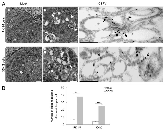
Figure 1. CSFV infection increases the formation of autophagosome-like vesicles. (A) PK-15 (a–c) and 3D4/2 (d–f) cells were mock-infected (a and d) or infected with CSFV (b and e) at an MOI of 1 for 48 h and studied by electron microscopy. White arrows indicate the structures with the characteristics of autophagosomes (b and e). The cells were also processed for IEM analysis. LC3 was visualized with specific antibodies and detected with a secondary antibody conjugated to 18-nm colloidal gold particles. LC3 protein immunogold labeling of CSFV-infected cells is shown in (c and f). The immunogold labeling localized to the infection-associated membranes is indicated by black arrows in the infected cells. Scale bar: 500 nm. (B) Quantification of the autophagosome-like vesicles per cell image. Average number of the vesicles in each cell was obtained from at least 10 cells undergoing each treatment. The data represent the mean ± SD of 3 independent experiments. Two-way ANOVA; ***P < 0.001.

An official website of the United States government
Here's how you know
Official websites use .gov
A
.gov website belongs to an official
government organization in the United States.
Secure .gov websites use HTTPS
A lock (
) or https:// means you've safely
connected to the .gov website. Share sensitive
information only on official, secure websites.
