Fig. 3.
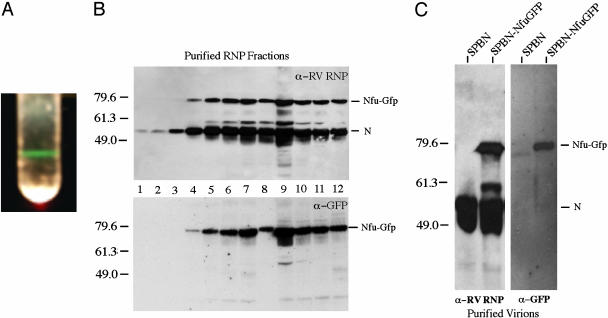
Characterization of recombinant RV expressing Nfu-GFP. (A) To analyze whether the recombinant Nfu-GFP protein is associated with RV RNPs, RNPs from SPBN-Nfu-GFP infected cells were purified by CsCl density centrifugation. A sharp band, which fluoresces green during exposure to light, can be seen. (B) Twelve fractions were collected from the CsCl gradient, dialyzed, and resolved by SDS/PAGE. Probing a Western blot with an RV N-specific antibody detected the highest concentration of RNPs in fraction 9, similar to the results seen for wild-type RNPs (not shown). In addition, a GFP-specific antibody detected equal amounts of Nfu-GFP and N in all fractions, further indicating incorporation of Nfu-GFP into RV RNPs. (C) Virions from BSR cells infected with SPBN or SPBN-Nfu-GFP were purified over 20% sucrose, separated by SDS/PAGE, and analyzed by Western blotting. The results indicate that the Nfu-GFP protein is incorporated into RV virions.

