Fig. 3.
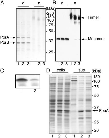
Protein and LPS profiles of wild-type (lanes 1), imp mutant (lanes 2), and lpxA mutant (lanes 3) bacteria. (A and B) Cell envelopes were analyzed by SDS/PAGE in denaturing (d) or seminative (n) conditions. Gels were stained with Coomassie blue (A) or were blotted and probed with anti-PorA antibody (B). (C) Equal amounts of proteinase K-treated cell lysates were subjected to Tricine-SDS/PAGE and stained with silver to visualize LPS. (D) Lanes labeled “cells” contain equal numbers of bacteria, based on OD. Lanes labeled “sup” contain equal volumes of culture supernatants precipitated with trichloroacetic acid. Samples were subjected to SDS/PAGE and were stained with Coomassie blue. Molecular size markers (in kDa) are indicated.

