
Figure 3. Crystal structure of Lys33-linked diUb.
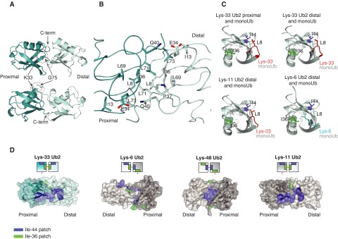
(A) The crystal structure of Lys33 diUb in two orientations. (B) Lys33 diUb is shown in ribbon and the residues at the interface are shown in stick representation. (C) Leu8 residue of Lys33-linked diUb contributes to Ile44 patch. Proximal Ub of Lys33 diUb and distal Ub of Lys33 diUb, Lys11 diUb (PDB 3NOB [10]) and Lys6 diUb (PDB 2XK5 [8]) were superposed with monoUb (PDB 1UBQ [41]) and coloured light cyan. The position of Leu8 (red), Ile36 (green) and Ile44 (blue) are indicated. As reference, the Leu8 of monoUb is not coloured differently. (D) A semi-transparent surface, coloured blue for Ile44 patch (Ile44, Leu8, His68 and Val70) and green for Ile36 patch (Ile36, Leu71 and Leu73) of diUb linked via Lys33, Lys6, Lys48 and Lys11 (PDB 2XK5, 3NOB, 1AAR [8,10,12]).