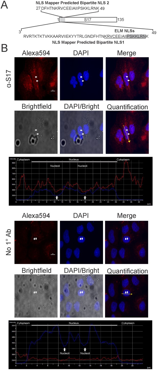
Fig 1. Schematic diagram and localization of human RPS17.

(A) Schematic depiction of RPS17 with a blowout showing the amino acids implicated via NLS mapper software. Eukaryotic linear motif (ELM NLSs) predictions are underlined for the bipartite NLS and shaded for the monopartite NLS. (B) Immunofluorescent staining of Huh7 human liver cells with anti-RPS17 antibody (Top) or with no primary antibody (Bottom) observed with confocal microscopy. Fluorescent intensity in DAPI and Alex594 channels was quantified along a yellow line through one representative cell (Quantification) from both anti-RPS17 antibody or no primary antibody stained cells showing RPS17 signal (red line) with respect to the cytoplasm, nucleus, and nucleolus (blue line). RPS17 localized predominantly to punctate spots within the nucleus as evidenced by the ALEXA594 staining overlaid with DAPI and brightfield images. There was minor nonspecific staining of the anti-mouse Alexa594 antibody within the cytoplasm but none detected in the nucleus suggesting the staining was specific. Small white arrows correspond to the same pair of nucleoli in each panel and were used for the representative quantification. Scale bars on Alexa594 images represent 10 μm.
