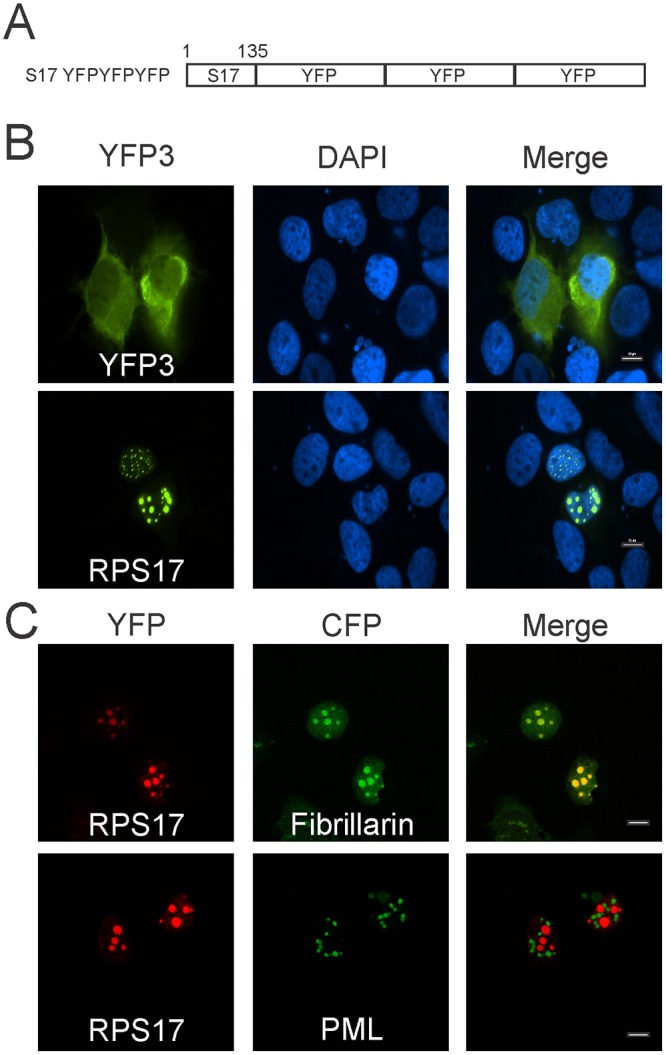
Fig 2. Subcellular localization of RPS17 attached to triple yellow fluorescent protein (eYFP).

(A) Schematic depiction of RPS17 as a carboxy-terminal fusion to RPS17. (B) RPS17 was capable of trafficking triple YFP to punctate spots within the nucleus as observed via confocal microscopy. Triple eYFP alone was predominantly cytoplasmic (Top) whereas RPS17 triple eYFP was in punctate spots within the nucleus (Bottom). (C) RPS17 triple eYFP is located within nucleoli. RPS17 triple eYFP colocalized with fibrillarin CFP (nucleolar marker) but not promyelocytic leukemia (PML) protein (PML body marker). Scale bars on merged images represent 10 μm.
