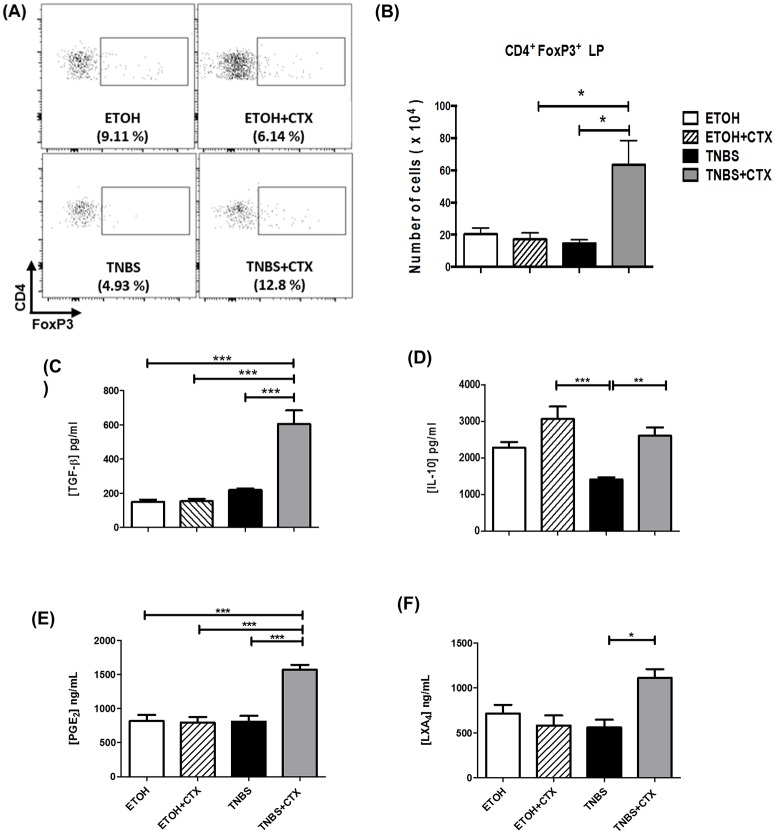
Fig 5. Analysis of CD4+FoxP3+ cells and anti-inflammatory mediators in TNBS-mice treated or not with CTX.
Cell suspensions were prepared from lamina propria of distinct experimental groups after 4 days of TNBS-induced colitis for the analysis of CD4+FoxP3+ cells by flow cytometry. The samples were prepared from a pool of cells from 4–5 animals/group performed in duplicate. The results are from 2 independent experiments. (A) Representative dot plots of CD4+FoxP3+ cells in the lamina propria obtained from distinct experimental groups. (B) Results of CD4+FoxP3+ cells expressed as a mean of the absolute number of cells in duplicate of 2 independent experiments ± SEM. Secretion of TGF-β (C) and IL-10 (D) in colonic tissue homogenates determined by ELISA. Production of PGE2 (E) and LXA4 (F) was performed by ELISA in colonic tissue homogenates. The results represent the mean obtained in individual mice/group ± SEM. * p<0.05, ** p<0.01 and *** p<0.001; (n = 4–5 animals/group).

