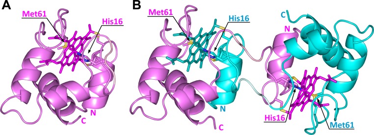
Fig 2. Crystal structures of monomeric and dimeric WT PA cyt c 551.
(A) Structure of monomeric WT PA cyt c 551 (PDB ID: 351C). (B) Structure of dimeric WT PA cyt c 551 solved in this study (pink and cyan, PDB ID: 3X39). The two protomers are depicted in pink and cyan, respectively. The hemes, Cys12, Cys15, His16, and Met61 are shown as stick models. The N- and C-termini are labeled as N and C, respectively. The hemes and Thr20–Met22 residues (hinge loop) are depicted in dark and pale colors, respectively. The sulfur atoms of the heme axial Met ligand and heme-linked Cys are shown in yellow, and the nitrogen atoms of the heme axial His ligand are shown in blue.

