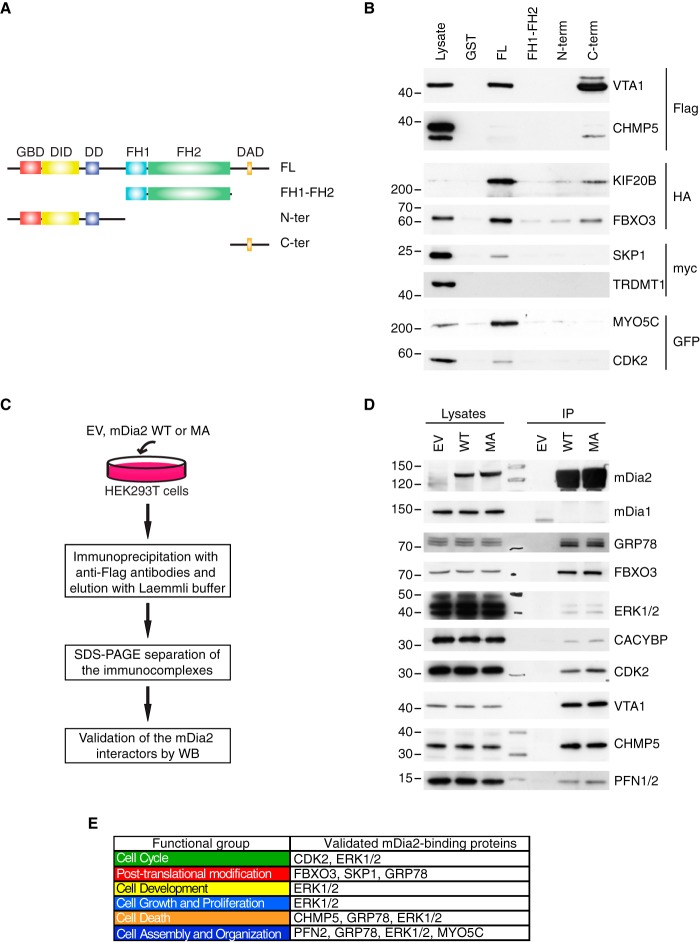
Fig. 2.
Validation of selected mDia2-interacting proteins. A, Schematic of the mDia2-deletion mutants. GST-mDia2 proteins used in the pull-down assays: full-length mDia2 = FL; Formin homology domain 1 and 2 (aa: 530–1033) = FH1-FH2; N terminus (aa: 1–530) = N-ter; C terminus (aa: 1033–1172) = C-ter. B, Validations by pull-down. Lysates obtained from 293T cells transfected with an epitope-tagged version of the candidate of interest (on the right) were incubated with immobilized GST-tagged, full-length mDia2, the mDia2-deletion mutants depicted in A, or GST as a negative control (on the top). Lysates (2%) and affinity-precipitated material were probed as indicated. C, Overview of the co-immunoprecipitation approach. The flowchart is as in supplemental Fig. S1D with the exception of the two following changes: (1) immunoprecipitation (IP) was carried out starting from 1 mg of total cell lysate, (2) Western blots were performed. D, Validations by co-immunoprecipitation. Endogenous proteins in the lysates (2%) and in the bound material were detected with specific antibodies (on the right). The expression of mDia2 was confirmed using anti-Flag antibodies. E, Validated functional groups and proteins. Functional groups are color-coded as in Fig. 1D. Proteins are listed according to their respective functional group. The absence of some validated proteins is because of the IPA annotation.

