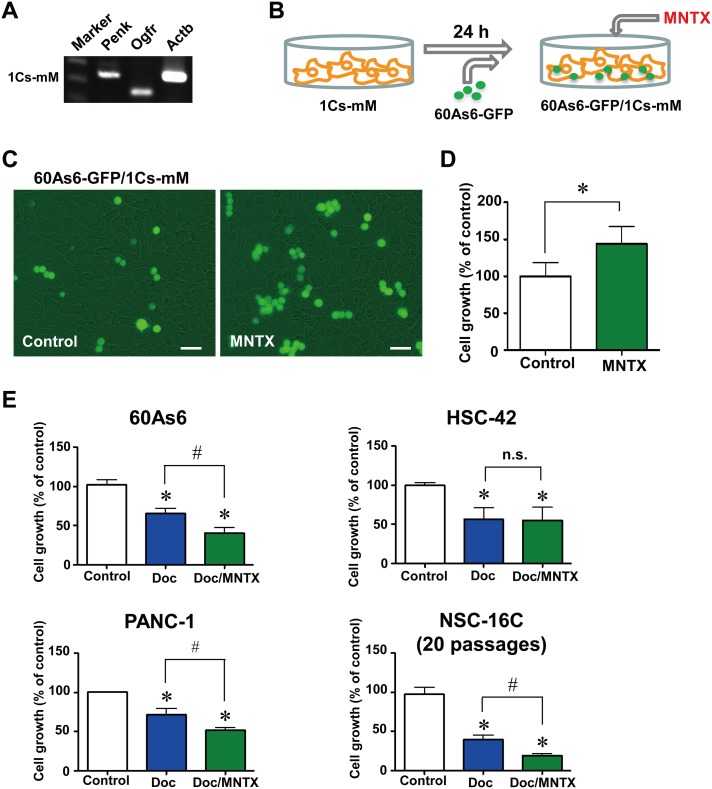
Fig 3. Blockade of OGF signaling by MNTX increased the growth of diffuse-type GC cells co-cultured with mesothelial cells.
A, RT-PCR analyses of Penk and Ogfr, in mouse mesothelial cells (1Cs-mM). B, schematic illustration of the system for co-culture of 60As6-GFP and 1Cs-mM. C, Growth of 60As6-GFP cells co-cultured with 1Cs-mM cells in the presence or absence of MNTX (10-5 M) for 72 h. Scale bar, 20 μm. D, the growth of 60As6-GFP cells was calculated (mean ± SD, n = 3 each, *p<0.05). E, growth of the diffuse-type GC cell line 60As6 cells, the intestinal-type GC cell line HSC-42 cells, the pancreatic cancer cell line PANC-1 cells, and primary cultured GC cells derived from the ascites of a patient NSC-16C cells treated with Doc (10-9 M) or a vehicle for 48 h, and subsequently treated with Doc, Doc/MNTX (10-6 M) or a vehicle for 48 h. Cells were counted with a hemacytometer (mean ± SD, n = 4 each, *p<0.05, vs. control, #p<0.05, Doc vs. Doc/MNTX).

