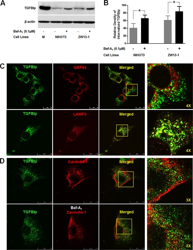
Fig 5. Internalized TGFBIp is transported to lysosomes.
A. NIH3T3 and ZW13-1 cells were pre-incubated for 60 min in the absence (-) or presence (+) of Baf-A1. After incubation, cells were incubated for 120 min in normal media containing TGFBIp and western blotting was performed for TGFBIp. B. Densitometric quantitation of the experimental results presented in A. A Student’s t-test was performed to determine the significance of differences between treatments with and without Baf-A1. Data analysis showed that the p-value was less than 0.05, indicating statistical significance. The experiment was repeated three times independently. C. Co-localization of TGFBIp with GRP94, ER marker, and Lamp-2, lysosome marker, in NIH3T3 cells. Cells were subjected to immunocytochemical staining as described in Materials and Methods. Areas of co-localization appear as yellow regions in the merged image. The boxed area in the third panel was magnified and is presented as the fourth panel. D. Co-localization of TGFBIp with caveolin-1 in the absence (upper panel) and presence (lower panel) of Baf-A1. NIH3T3 cells were grown on glass slides and treated with vehicle or Baf-A1 (0.1 μM) for 60 min before incubation for 30 min at 4°C in medium containing ~1 μg/mL TGFBIp. The cells were subjected to immunocytochemical staining as described in Materials and Methods. The boxed area in the third panel was magnified and is presented as the fourth panel.

