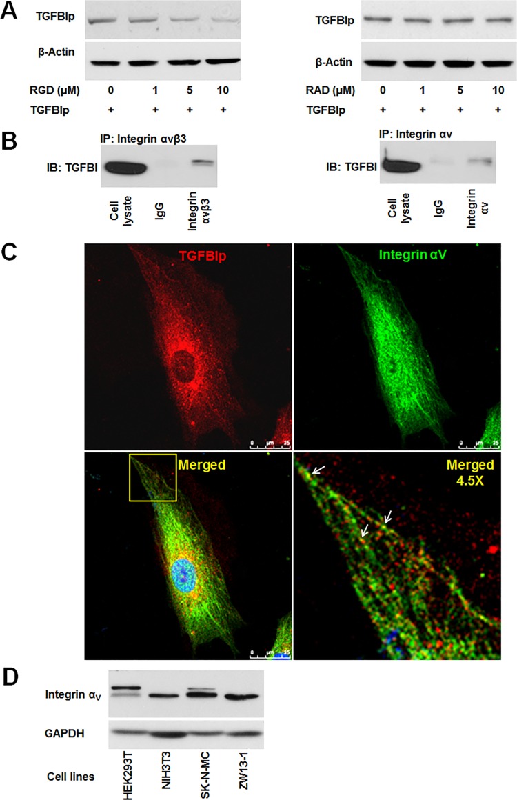
Fig 6. Integrin-dependent endocytosis of TGFBIp in corneal fibroblasts.
A. Endocytosis of TGFBIp was blocked by RGD peptide in a dose-dependent manner. Corneal fibroblasts were pre-incubated for 30 min in the absence (lane 1) or presence (lanes 2–4) of RGD or RAD peptides. TGFBIp (~1 μg/mL) was added to the medium and the cells were incubated for 120 min at 37°C. TGFBIp levels were measured by western blot analysis. B. TGFBIp interacts with integrin αVβ3 and αV. Cells were lysed with RIPA buffer and the lysate was immunoprecipitated with anti-integrin αVβ3 (left-hand panel) or anti-integrin αV (right-hand panel) antibody as indicated. Immunoprecipitates were resolved on 10% SDS-PAGE gels and immunoblotted with anti-TGFBIp polyclonal antibody. C. Co-localization of integrin αV with TGFBIp was visualized by confocal immunofluorescence microscopy. The merged images show TGFBIp as red, integrin αV as green, and areas of co-localization as yellow. The boxed area in the lower left-hand panel was magnified and is presented as the lower right-hand panel. Arrows identify regions of TGFBIp and integrin αV co-localization. Scale bars, 5 μm. D. Western blot analysis of HEK293T, NIH3T3, SK-N-MC, and ZW13-1 cell lines with monoclonal antibody against integrin αV. GAPDH was used as a loading control.

