Fig. 12.
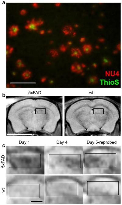
Diagnostic assays for Aβ oligomer levels provide AβO-relevant MRI signals in brain. a Sagittal brain sections, 50 µm thick, from 8-month-old 5xFAD and wt mice were probed with 568-NU4 and counterstained with Thioflavin S. Image shows a 5xFAD cortical region stained with both NU4 (red) and thioflavin S (green). NU4 labeling is more abundant than the ThioS staining. Findings demonstrate that NU4 labeling is often associated with, yet distinct from, amyloid plaques. Scale 25 µm. b In vivo imaging of NU4MNS distribution in live mice 4 h after intranasal inoculation shows labeling by the probe in the hippocampal region of the Tg mice, but not the wt controls. Scale bar 5 mm. c Higher magnification of the hippocampal region of the Tg and wt mice shows probe distribution 4 h after inoculation, the changes in distribution 96 h later, and the distribution of the target after re-administering the probe on Day 5. Data suggest that non oligomer-associated probe is clearing the brain. Scale bar 1 mm. Adapted from Viola et al. [174]

