
Fig. 8.
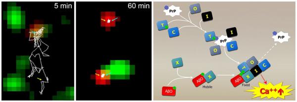
Single molecule trafficking shows AβOs stop diffusion of mGluR5 and “highjack” membrane proteins that can lead to elevated Ca2+. Left panels Dual-color single-particle tracking was used to monitor mGluR5 (red) and biotin-AβO (green) diffusion at syn- apses over time. Following the tracings of mGluR5, mGluR5 diffuses together with an AβO (5 min) outside synapses before both become stabilized at a synaptic site (60 min). Adapted from Renner et al. [143]. Right Clustering of membrane proteins, possibly involving PrPc, leads to AβO binding recruitment and membrane receptor reorganization that instigates toxic signaling. AβO binding to an unidentified receptor, X, and the recruitment of effector protein co-receptors leads to hyperactive Ca2+ signaling and downstream toxicity