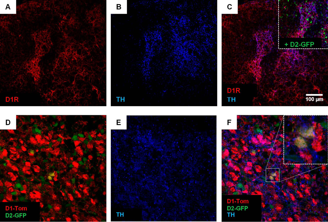
Figure 3. Dopamine receptor co-expression occurs within the patch compartment of the neonatal striatum.
(A, B, C) A representative photomicrograph showing double staining for native D1R (A) and tyrosine hydroxylase (TH,B), a marker for the patch compartment in neonatal striatum. The almost complete overlap between D1R and TH signal is shown in (C), suggesting that clusters of D1R expression are endemic to the patch compartment. For cross-methodological comparison, the inset in (C) illustrates the higher fluorescence intensity of D2-GFP expressing cells within TH-positive clusters than outside these regions, which agrees with our finding of higher native D2R expression within D1R-positive patches (Figure 1). (D, E, F) A representative photomicrograph showing that cells with high D1-Tom and/or D2-GFP levels (D) are clustered within TH-positive regions (E,F), suggesting that these clusters are patch-specific. Inset in (F) illustrates an example of dual-expressing D1-Tom/D2-GFP cells within the patch compartment.

