Figure 1.
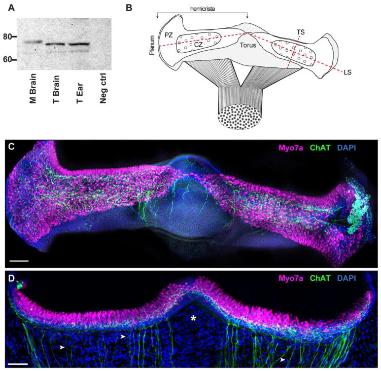
The expression of ChAT in turtle canal cristae. A. Specificity of the ChAT antibody (AB144P) was confirmed via western blotting of turtle and mouse tissues. Both tissues showed ChAT immunoreactivity within the appropriate range of molecular weights. Lanes (left to right): 1, Mouse brain; 2, Turtle brain; 3, Turtle ear; 4, negative control. B. Diagram of the intact turtle posterior crista modeled after the immunostained whole organ micrograph shown in panel C. The canal nerve bifurcates to innervate two triangular-shaped hemicrista that extend from the planum to the nonsensory torus. Each hemicrista consists of a central zone (CZ) and peripheral zone (PZ). Dashed lines indicate planes of sectioning used in this study (LS – longitudinal section; TS – transverse section). C. Apotome image projection of a whole crista preparation of the turtle posterior crista where cholinergic fibers (green) and hair cells (magenta) were labeled with antibodies to ChAT and Myo7A, respectively. Nuclear staining with DAPI is shown in blue. D. Confocal image projection of a longitudinal section of the turtle posterior crista stained for ChAT (green) and Myo7A (magenta). Intense DAPI staining (blue) below hair cells demarcates supporting cell layer. Arrowheads identify single ChAT-positive fibers and asterisk demarcates stromal region devoid of efferent fibers. Scale bars = 50 μm.

