Figure 2.
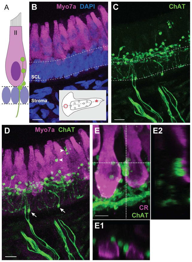
ChAT-positive fibers give rise to numerous spherical varicosities in the hair cell layer. A. Illustration depicting common locations of efferent terminals on type II hair cells. Supporting cell nuclei fill the space beneath the hair cell layer. B. Confocal image projection of a longitudinal section of the turtle crista where type II hair cells near the torus (asterisk, inset) were stained with antibodies to Myo7A (magenta). Dense DAPI staining (blue) emphasizes the supporting cell layer (SCL, dashed lines). C. Efferent fibers and associated varicosities were labeled with antibodies to ChAT (green) in the same section shown in panel B. D. Combined image details the arrangement of ChAT-positive varicosities along type II hair cells. Arrows indicate ChAT-positive varicosities located well below the hair cell layer. Arrowheads identify more apical ChAT-positive varicosities. E. Confocal image projection of a longitudinal section of the turtle crista showing type II hair cells near the planum stained with antibodies to calretinin (magenta). ChAT-positive varicosities (green) populate the basal pole. E1, E2. Orthogonal views, generated along the dashed lines, indicate the relative position of ChAT-positive varicosities within the Z-plane. Scale bar = 10 μm in B–D, 5 μm in E.

