Figure 4.
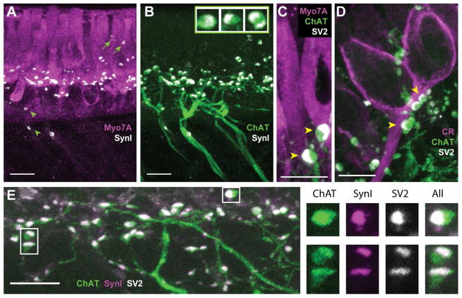
The synaptic vesicle proteins synapsin I and SV2 colocalize with ChAT in efferent varicosities of the turtle crista. A, B. Confocal image projection was generated from a longitudinal section of the turtle crista stained with antibodies to Myo7A (magenta), synapsin I (SynI, white), and ChAT (green). A. Antibodies to SynI labeled numerous puncta along the basal end of type II hair cells near the torus. A few SynI-positive puncta were also located the above hair cell nuclei (arrows) and below the hair cell layer (arrowheads). B. Most of the puncta stained with antibodies to SynI appeared restricted to ChAT-positive varicosities. Inset: Zoomed images showing colocalization and distribution of ChAT (green) and synI (white) within three efferent varicosities. C, D. Confocal image projections were generated from transverse sections of the turtle crista previously stained with antibodies to Myo7A (magenta in panel C) or CR (magenta in panel D) with ChAT (green), and SV2 (white). Several efferent varicosities cluster along the base of a type II hair cell near the torus (panel C) and calyx afferent from the central zone (panel D). E. A confocal image projection of efferent fibers and varicosities near the torus taken from a longitudinal section of a turtle crista stained for ChAT (green), SynI, (magenta), and SV2 (white). Right panels show zoomed images of three efferent varicosities (boxes, panel D) showing the colocalization and distribution of ChAT, synI, and SV2. Scale bar = 10 μm in all panels.

