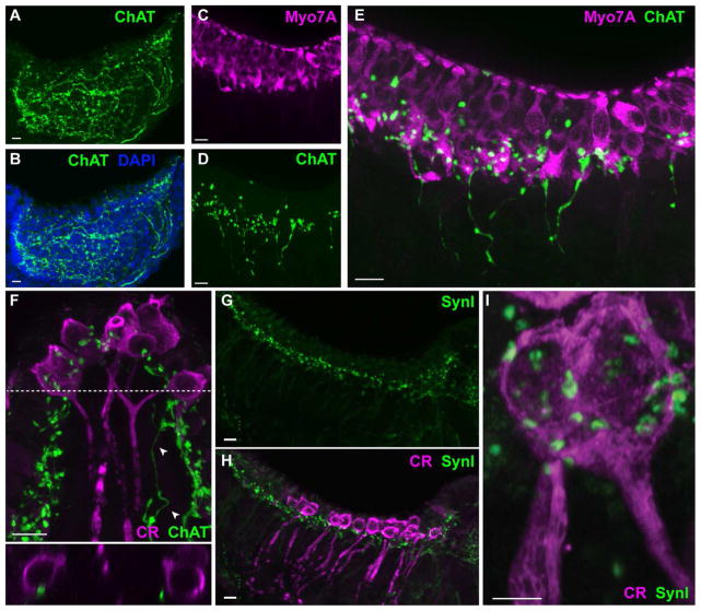
Figure 7.
ChAT is a marker of vestibular efferents in mouse cristae. Confocal image projections were generated from either longitudinal (panels A–E, G–I) or transverse (panel F) sections of the mouse crista. A, B. Antibodies to ChAT (green) labeled efferent fibers and varicosities across the crista. DAPI staining (blue) defines the tissue boundaries. C. Hair cells in the mouse crista were stained with antibodies to Myo7A (magenta). D. Efferent fibers and associated varicosities were labeled with antibodies to ChAT (green) in the same section shown in panel C. E. Combined image from panels C and D shows ChAT-positive varicosities scattered along the hair cell layer. F. Calyx-bearing afferents in the central zone of the mouse crista stained with antibodies against calretinin (magenta). ChAT-positive varicosities (green) are distributed across the neuroepithelium with several located in close apposition to calyx endings. A ChAT-positive fiber can be seen on the right (arrowheads). Bottom panel: Orthogonal view along dashed line indicates the proximity of varicosities to the outer face of two calyx endings. G, H. Confocal image projections of mouse hemicrista stained with antibodies to SynI (green) and calretinin (magenta), respectively. I. An enlarged image of a calretinin-positive calyx (magenta) shows the location of several SynI-positive puncta. Scale bar = 10 μm except Panel I which is 5 μm.

