
Figure 1.
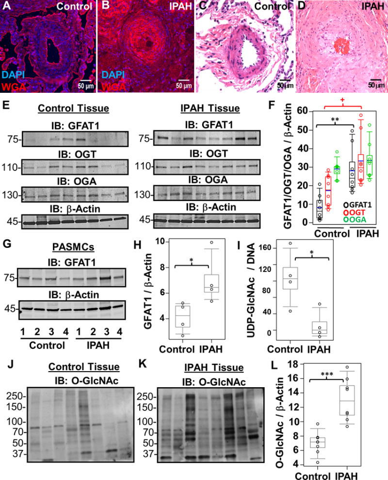
Increased hexosamine biosynthesis results in global glycosylation changes in IPAH. Representative WGA lectin (A and B) and H&E (C and D) stains of a control (A and C) and IPAH (B and D) occluded vessel within a plexogenic lesion from serial sections of the same lung tissue. (E) Characteristic Immunoblots of GFAT1, OGT, and OGA from multiple control and IPAH human tissues isolated from explant lungs (n=8). Lysates were normalized to total protein and β-Actin was used as a loading control. (F) Densitometry quantitation of each protein compared to β-Actin. (G) GFAT1 was also probed in multiple control and IPAH human PASMCs isolated from pulmonary arteries (n= 4) and quantitated (H). (I) UDP-GlcNAc pools were isolated (see Methods) and analyzed from multiple control and IPAH PASMCs (n=3). UDP-GlcNAc levels were normalized to total DNA concentration. In addition, total O-GlcNAc levels were analyzed in control (J) and IPAH (K) lung tissue. The global protein O-GlcNac levels were normalized to β-Actin (E) and quantified (L). As a reactivity control, the O-GlcNAc antibody was pre-treated with 250 mM GlcNAc before primary incubation (Supplemental Figure 3). The p-values were calculated based on a Wilcoxon test (see Materials and Methods) determined from the independent experiments (+, p=0.027; *, p<0.05; **, p<0.01; and ***, p<0.001).