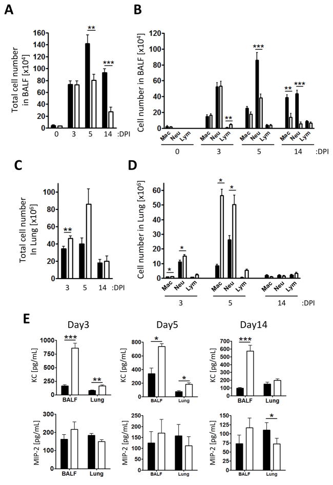
Figure 2. Wsh mice displayed reduced inflammatory cell infiltration into the airspace compared to WT.
WT (closed bar) and Wsh (open bar) mice were infected with 2×10⁶ Cpn intratracheally. BALF was collected from euthanized mice on 3, 5, and 14 days post Cpn propagation. Total (A) and differential (B) cell counts in BALF. Cell types in BALF were determined by modified Wright-Giemza staining. (C and D) Single cells from lung were collected through enzymatic procedure and analyzed by flow cytometry. (E) Chemokine concentrations in BALF and the lung homogenates were determined by ELISA. This experiment was performed 3 times and the data was pooled for a total n=7 (WT and Wsh) day 0, n=10 (WT) and n=8 (Wsh) day 3, n=9 (WT and Wsh) day 5, and n=7 (WT) n=9 (Wsh) day 14. Data are mean ±SD. Significance of differences was determined by Mann-Whitney. *P<0.05, **P<0.005, ***P<0.001.

