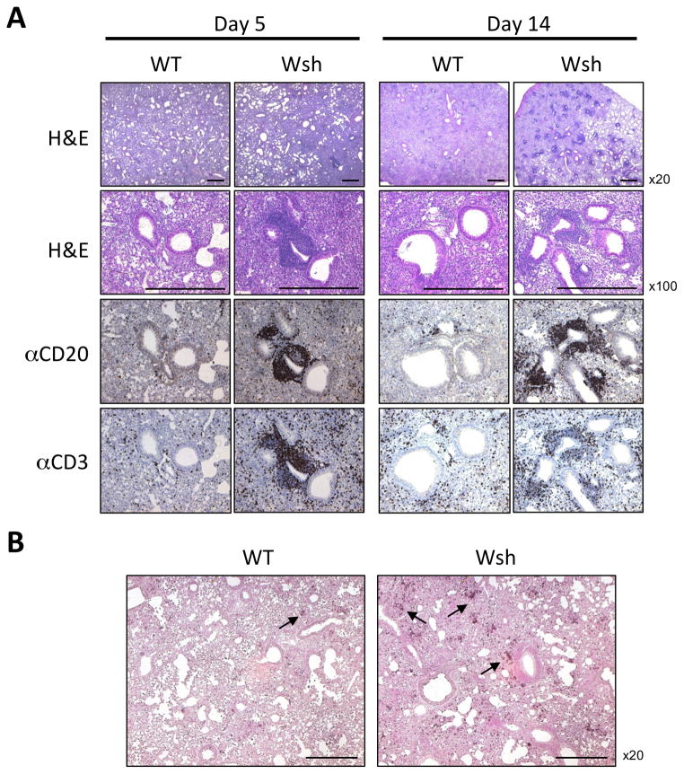
Figure 6. Wsh mice displayed patchy inflammation and accumulated T and B cells perivascularly and accumulated neutrophils in the lung during Cpn-induced lung inflammation.
WT and Wsh mice were infected with Cpn intratracheally. Lungs were harvested for HE staining and immunohistochemistry for B cells and T cells on day 5 and day 14 (A) and neutrophils on day 5 (B). This experiment was performed 3 times and the data was pooled for a total n=7 (WT and Wsh) day 5, and n=7 (WT) n=10 (Wsh) day 14. Bar in the picture indicated 0.5mm.

