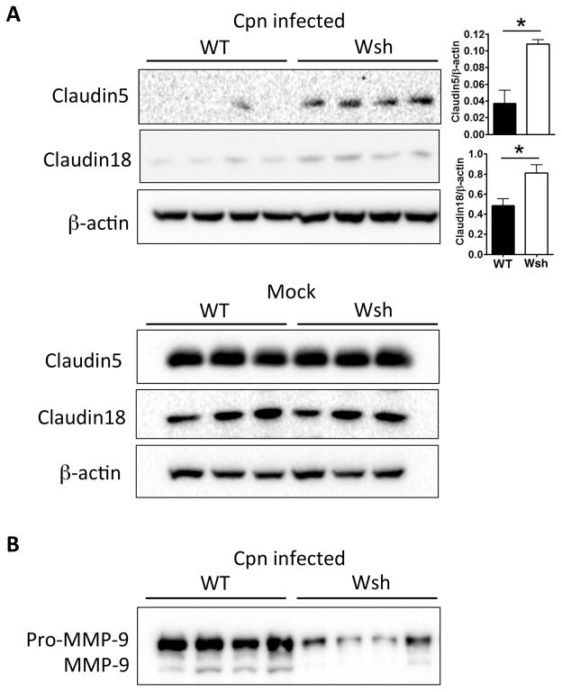
Figure 7. Cell-cell tight junction molecule degradation was reduced in Wsh lung compared to WT during Cpn lung infection.
WT and Wsh mice were infected with 2×10⁶ IFU Cpn intratracheally and sacrificed on day 5. (A) Lung cell lysates were separated by SDS-PAGE and immunoblotting was performed with indicated antibodies. The representative westerns are shown to analyze relative amounts of claudin 5 and 18 in the lung from naïve and Cpn-infected WT and Wsh mice. (B) BALF was separated by SDS-PAGE and immunoblotting was performed with anti-MMP-9 Ab to determined secreted MMP-9 in BALF. For A, this experiment was performed 3 times n=5 (WT) n=6 (Wsh). For A & B, the experiment was performed twice n=4 (WT) n=4 (Wsh). Data shown is a representative experiment. Data are mean ±SD. Significance of differences was determined by Mann-Whitney. *P<0.05.

