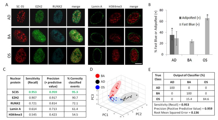
Fig. 2. SC-35 organization as a screen to parse stem cell differentiation.
hMSCs were cultured in BA, AD, or OS media and (A) fixed and immunolabeled for a panel of either gene-modifying (H3K4me3, EZH2, RUNX2) or mechanoresponsive (Lamin A, SC-35) proteins 3 days post-plating. (B) hMSCs cultured in BA, AD, or OS media for 14 days were stained for intracellular triglycerides (AdipoRed) and alkaline phosphatase (ALP) to identify % of adipocytes and osteoblasts, respectively. (C) 3 day analysis of nuclear proteins yields differences in sensitivity, precision, and % of correctly classified events, and (D) PCA plot of SC-35 reporter shows that the three conditions cluster into three distinct groupings (blue = OS, black = AD, red = BA). (E) Decision tree analysis of PCA-derived principal components show that hMSCs parse well (> 95%). Scale bar = 5 μm. * indicates statistical significance (p < 0.01). Sample size ranged from 13 to 15 cells per nuclear reporter per growth factor treatment condition.

