
Figure 7.
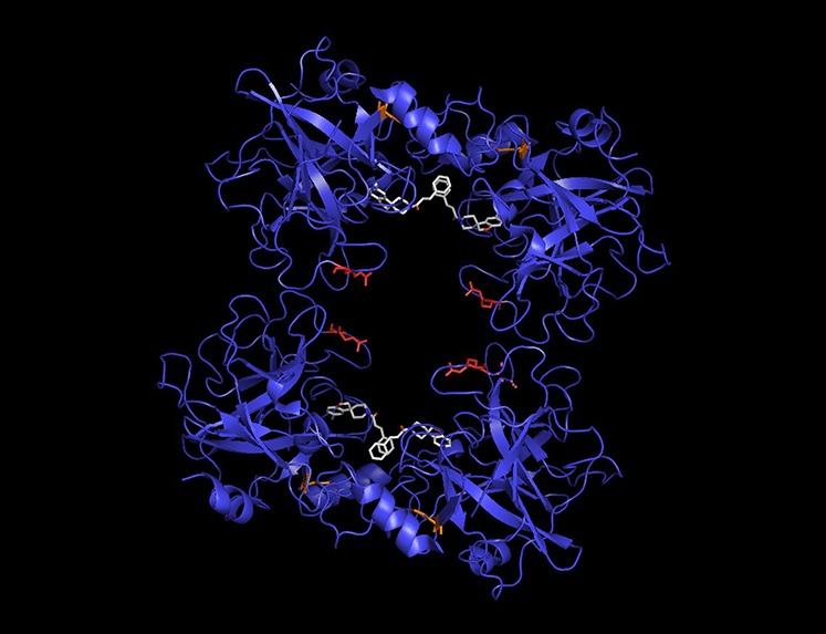
Example: a MOSAIC-specific PSS in Tryptase Alpha/Beta 1 (TPSAB1). The tetrameric TPSAB1 structure is shown with positively selected sites highlighted. The site detected by component methods and by MOSAIC is colored orange, whereas the MOSAIC-specific PSS is featured in red. A bound inhibitor (white) pinpoints the active site of the enzyme.