
Figure 4.
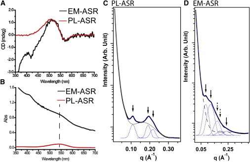
(A) Visible range CD and (B) absorption spectra of PL-ASR and EM-ASR. In (A), the bilobal shape is clearly present in both PL-ASR and EM-ASR, indicating the presence of trimers. SAXS spectra of (C) PL-ASR and (D) EM-ASR. Bragg diffraction peaks in the SAXS spectrum of PL-ASR indicate the presence of an ordered two-dimensional hexagonal lattice, while the peaks present in the EM-ASR spectrum indicate the presence of a two-dimensional tetragonal lattice. See text for details. To see this figure in color, go online