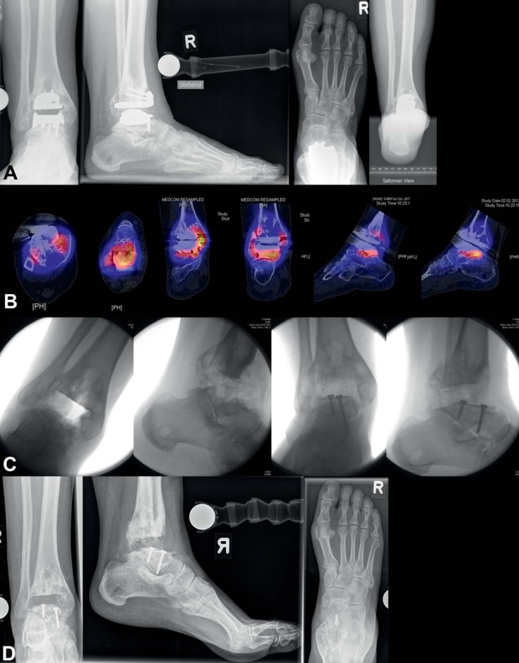
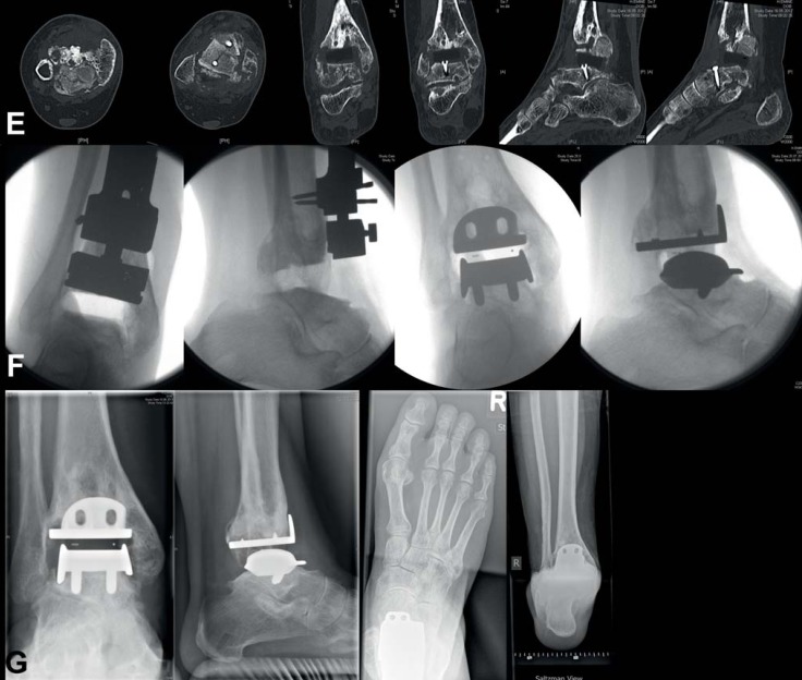
eFigure 2.
Aseptic loosening of total ankle replacement 8 years after initial surgery: a) conventional X-rays in standing position show borders of loosening around both implant components, tibial and talar; b) SPECT-CT shows high metabolic activity in the areas around both implant components; c) implant components have been removed, on talar side osseous reconstruction was performed using autograft taken from iliac crest; d) and e) conventional X-rays and CT show good bone consolidation of autograft from iliac crest on the talar side 4 months postoperatively; f) fixation screws on talar side have been removed, HINTEGRA implant (revision components on talar side) were implanted; g) conventional X-rays in standing position show good osseointegration of implant components 6 months postoperatively


