
Figure 2.
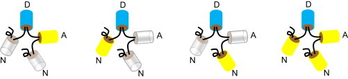
Schematic of the cytoplasmic FRET standards. A donor (D, Cerulean), an acceptor (A, Venus), and two nonfluorescent proteins (N, Amber) are joined by amino-acid linkers depicted (by curved lines connecting D, A, and N (28)). To see this figure in color, go online.