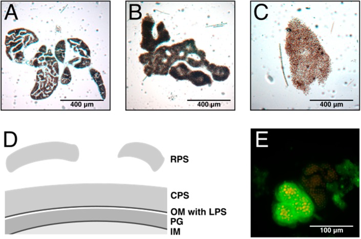
Figure 1.
EPS in Microcystis. Light micrographs of characteristic morphotypes of (A) M. wesenbergii. (B) M. aeruginosa and (C) Microcystis sp. The colony morphology is determined by the mucilage embedding the cells. (D) The exopolysaccharides (EPS) are further classified as O-antigens of lipopolysaccharides (LPS) anchored in the outer membrane (OM), capsular polysaccharides (CPS), which are associated with the cell surface, and released polysaccharides (RPS), which are secreted to the culture medium without attachment to the producing cells. PG, peptidoglycan; IM, inner membrane. (E) The fluorescein isothiocyanate-labelled lectin, microvirin, bound to a Microcystis colony. Selective binding of MVN shows different exopolysaccharide composition in identical Microcystis morphotypes.

