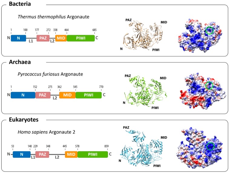
Figure 1.
Overall architecture of Argonaute from the three domains of life. The domain composition (left) and structures (middle) of the bacterial (based on Thermus thermophilus, PDB: 3DLH), the archaeal (based on Pyrococcus furiosus, PDB: 1U04) and the eukaryotic (based on human Argonaute 2, PDB: 4EI3) Argonaute reveal an evolutionarily conserved architecture. Differences can be found in the surface charge distribution of Argonaute proteins (negatively-charged surfaces in red; positively-charged surfaces in blue). The binding pocket for the 5'-end of the guide in the MID domain is highlighted with a green circle.

