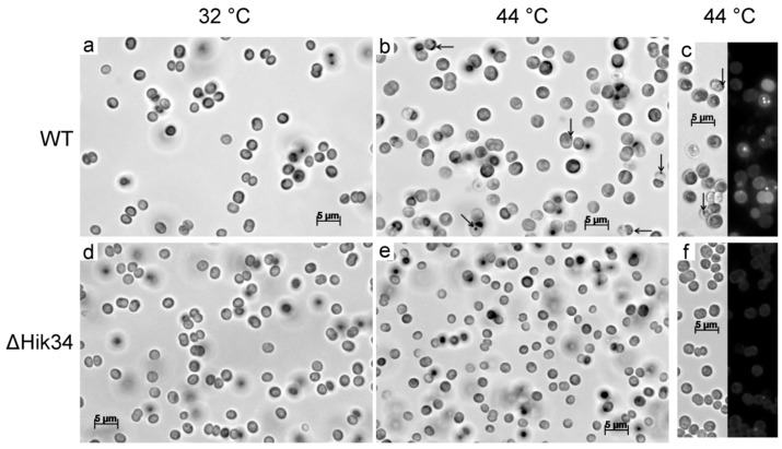
Figure 5.
Light and fluorescent microscopy pictures of wild-type (a, b, c) and ΔHik34 mutant (d, e, f) cells. Cells were grown 24 h at 32 °C (a, d) and thereafter incubated 24 h at 44 °C (b, c, e, f). Images of cells stained with Nile Red (c, f) were received from two channels—bright field (left panel) and fluorescence (right panel). Arrows point to unidentified inclusions. Cells were analyzed in 3 independent experiments: typical microscopic photos of cells are shown.

